
 |
|
Ronaldo Hueb Baroni1,a; Tufik Bauab Jr.2,b; Leonardo Kayat Bittencourt3,c; Giuseppe D’Ippolito4,d; Suzan Menasce Goldman4,e; Guilherme Hohgraefe Neto5,f; Adonis Manzella6,g; Antonio José Rocha7,h; Luis Augusto Sonoda8,i; Fabio Seichi Takeda9,j
– International guidelines and consensus state that macrocyclic GBCAs are safer than linear GBCAs in terms of brain deposition. Do you agree that macrocyclic GBCAs have a lower risk of brain deposition?Seven participants answered “yes” and three answered “no”. Two participants said that they had not conducted studies to make such a statement and one said that he/she did not have enough experience with brain MRI.
– The European Medicines Agency has restricted the use of linear GBCAs, but these are widely used in Brazil. What should be done in Brazil?Six participants answered that linear GBCAs should no longer be used, except in cases of organ-specific contrast agents. Four answered that linear GBCAs should continue to be used as a secondary approach.
– Is gadolinium deposition in body tissues a concern among Brazilian radiologists?Six participants answered “yes” and four answered “no”. However, the entire panel is concerned about this potential risk, according to the comments associated with the responses to this question:
• Radiologists outside Brazil care more about this potential risk. • It should be a cause for concern, but the lack of guidelines and long-term data make it a non-priority. One participant observed that this fact gets more attention outside Brazil. • The assistant physicians are not concerned with this possibility, or they are unaware of it. – What should be done to raise the awareness of the importance of the issue of gadolinium deposition and to decrease the long-term risks?The participants could choose any alternative that was applicable. The answers are summarized in Figure 1.
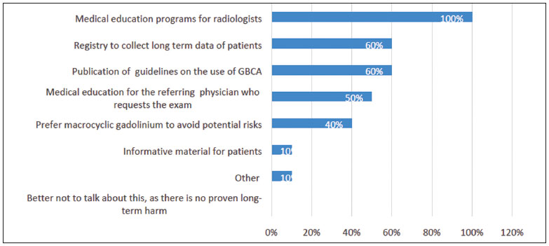 Figure 1. Recommendations to raise awareness of gadolinium deposition and decrease the potential associated long-term risks.
– Is there a difference in the image resolution obtained on using 1.0 M (1 mol/L) gadolinium and that obtained using 0.5 M gadolinium?Two participants answered “yes”, four answered “no”, and four answered “other”. The “other” responses were explained as follows:
• In some cases, 1.0 M gadolinium results in superior image resolution. • It depends on the volume to be injected. • There are differences between gadobutrol and gadoteric acid. • Theoretically, 1.0 M gadolinium could result in a superior image. However, I do not see it in my practice. – Do you think that, ideally, we should reduce the volume of 1.0 M GBCA used, in order to adjust it to the lowest effective dosage that enables the obtainment of a good-quality image?Nine participants answered “yes, we should ideally reduce the injected volume”. One participant commented that this reduction should be done only in case of renal impairment. Overall, they stated that their opinion was based on an ideal situation, but evidence is needed to make such statement. The same question was posed regarding the volume of 0.5 M GBCA. Again, the majority (eight participants) agreed that ideally the volume should be reduced whenever possible. One participant reminded that image quality correlates with dosage, but in cases of exams of the pancreas and liver, it is not possible to reduce the dosage. In this question, we asked the radiologists whether the volume should be ideally reduced. In the next question, we asked if it was feasible.
– Is it possible to use 1.0 M GBCA at dosages/volumes lower than 0.1 mL/kg and still obtain an image with good accuracy for diagnosis?We received seven “yes” and three “no” responses. One participant confirmed that he/she had obtained good results with lower dosages according to personal experience; another observed that reducing the dose would be adequate in some cases, but not for the pancreas and liver.
– Does the minimal volume to be injected depend on the scanner and protocol?Seven participants answered “yes”, two answered “no”, and one said that it also depended on the clinical condition of the patient.
– What is your perception of the image quality obtained with 1.0 M GBCA in dosages lower than 0.1 mL/kg?Seven participants answered that they perceived the image quality to be at least as good as that obtained with the dosage of 0.1 mL/kg. The words used for these answers were “excellent”, “satisfactory”, “good”, and “the same”. This was not a multiple-choice question. One participant answered that the quality was worse, and two said that they did not have experience with this dosage. We had one more discussion about this perception on the second round of questions; overall, the participants reaffirmed that this was a perception, and more evidence is needed before confirming that a lower dose is efficient. Some participants were concerned that a dosage below 0.1 mL/kg was an “off-label” dosage. However, the prescribing information states that 0.1 mL/kg is “usually sufficient”, but does not state that is mandatory to use this dosage(11). Six participants reported that they had experience with a lower dosage of 1.0 M GBCA.
– Is it possible to make an accurate diagnosis using lower dosages of 0.5 M GBCA? What is your opinion?Five participants answered that it was possible to make a good diagnosis with a lower dosage of 0.5 M GBCA. Those who disagreed justified their responses with a lack of experience with a lower dosage and a lack of evidence.
– Considering the last reports of brain deposition, although we do not know the long-term effects of gadolinium deposition in the brain, do you think it is a concern that could justify the use of a lower volume of 1.0 M GBCA?Nine answered “yes”.
– In your opinion, what would be the minimal dosage of 1.0 M GBCA and 0.5 M GBCA that would allow for an accurate diagnosis?Four participants gave their opinion on a minimal dosage below 0.1 mL/kg for 1.0 M GBCA and 0.2 mL/kg for 0.5 M GBCA. For 1.0 M GBCA, one suggested a dosage of 0.035 mL/kg and another participant suggested a dosage of 0.06 mL/kg. For 0.5 M GBCA, one participant suggested a dosage of 0.15 mL/kg. One participant estimated an approximately 20% reduction and another participant mentioned that the experimental use of deep learning would allow a reduction to 10% of the current dosage. The participants also said that evidence is necessary before any recommendation, and it is important to consider the body region that is being examined.
– Do you believe that the lower injected volume decreases the risk for acute adverse reactions (e.g., allergic reactions)?Nine participants answered “no” to this question, and there was an agreement that the acute reactions do not depend on the dosage. However, one participant observed that although the dosage is not a risk factor for acute reactions, a lower dosage might be important to prevent nephrogenic systemic fibrosis and gadolinium deposition in tissues.
– Are macrocyclic GBCAs (0.5 M and 1.0 M) safe to be used in children, including those younger than 2 years in age and neonates?Seven participants agreed that macrocyclic GBCAs are safe for use in children, and three participants made observations agreeing with the safety of GBCAs. One mentioned a lack of experience with children, and two others recommended GBCA use only if extremely necessary, as its approval for use in children younger than 1 year in age is more recent and the urinary system in such children is immature.
– Is it necessary to adjust the dosage/volume (mL/kg) in children?Nine participants answered “no” to this question.
– Are macrocyclic agents safer than linear agents, in any age group?Eight participants answered “yes”, and two answered “no”. Two participants observed that macrocyclic agents are safer in terms of deposition in the brain and other tissues, but not in terms of acute adverse reactions.
– GBCAs should not be used in patients with severe renal impairment (glomerular filtration rate < 30 mL/min).Although seven participants agreed with this statement and three disagreed, the participants observed that it could be done if necessary, as justified by the clinical need. In such cases, a macrocyclic agent should be used.
– What should be done when the patient has impaired renal function, but this impairment is not severe (glomerular filtration rate > 30 mL/min)?In this question, multiple choices were accepted. The answers are summarized in Figure 2. The “other” answers were comments added to the alternatives:
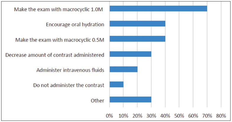 Figure 2. Management of patients with mild to moderate renal impairment.
• Two participants added: “It depends on the creatinine clearance”. • One participant commented that he/she would first check for the real need of contrast. – In your experience, do you think there is an anatomical region where a dosage/volume of 1.0 M macrocyclic GBCA lower than 0.1 mL/kg could provide an image with good accuracy?Six answered “do not know”, and four gave their opinion, as follows:
• Possibly any region, including the heart. • Pelvis, abdomen, cranial and joints. • Musculoskeletal. • Brain. – In your experience, are there differences between 1.0 M GBCA and 0.5 M GBCA in terms of artifacts?Only one participant observed differences in the presence of artifacts, observing that artifacts occur with gadobutrol in the pancreas, liver, and kidneys. Another panelist stated that it is theoretically possible to have artifacts with gadobutrol owing to its higher concentration; however, he/she did not observe it in clinical practice.
– Does the use of high-relaxivity contrast agents allow for sequences with better spatial resolution?Half of our panel agreed that higher relaxivity allows for better spatial resolution. One participant observed that the statement is correct if the aim is to investigate whether the production of more signal can aid in the detection of lesions. One participant reminded that in MRI, the concept of spatial resolution is related to the voxel size, which is a consequence of the slice thickness, field of view, and matrix size.
– In a non-emergency setting, would you administer GBCAs to a pregnant patient or to a patient with a suspicion of pregnancy?Multiple choices were accepted. The answers are summarized in Figure 3.
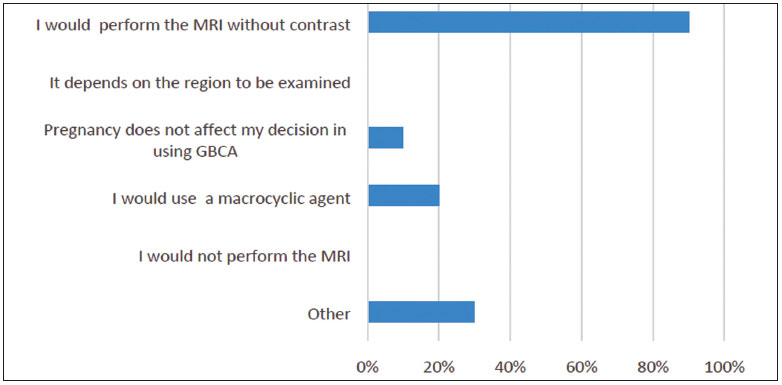 Figure 3. Use of GBCAs in a pregnant patient in a non-emergency setting.
– Would you administer GBCA to a woman who is breastfeeding?Multiple choices were accepted. Results are summarized in Figure 4.
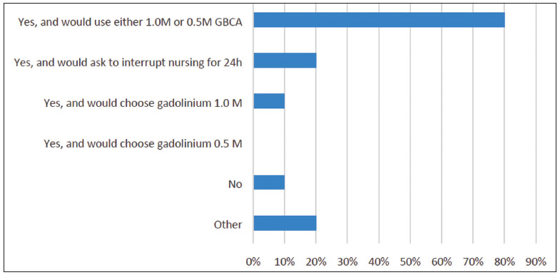 Figure 4. Use of GBCAs in women who are breastfeeding.
– Which criteria should be adopted for the diagnosis of acute renal insufficiency in clinical practice?Eight participants answered that the European Society of Urogenital Radiology criteria should be used, one participant preferred the Kidney Disease Improving Global Outcomes criteria, and one participant answered “both”. DISCUSSION Radiologists have long been using GBCAs in their daily practice, often without clear recommendations or evidence regarding the differences among the agents. Although some studies suggest that linear GBCAs deposit more gadolinium in the brain than macrocyclic GBCAs(6,12–14), linear GBCAs are still widely used in Brazil. Our panel tended to agree that the use of linear agents should be restricted to organ-specific exams (60% agreement) or as a secondary approach (40% agreement). There are also differences among macrocyclic agents. Half of our panel believed that it is possible to use a lower volume of 1.0 M GBCA to produce good-quality images, and nine experts (90%) believe the injected volume should be ideally reduced, in order to avoid the potential risks of the gadolinium deposition in the brain, bones, and skin. However, despite the experiences reported by members of the panel, the lack of controlled studies using lower volumes of gadolinium, either 0.5 M or 1.0 M, is a concern. The panelists suggested that clinical studies with lower volumes of gadolinium are necessary. The panel discussed issues related to pregnancy, breastfeeding, infants, and renal impairment; there were concerns regarding the potential unknown impact of gadolinium deposition and the lack of evidence to support several decisions. All experts agreed that a medical education program for specialists could raise awareness of the issues related to clinical decisions and long-term outcomes. The strength of this Delphi panel was the undeniable expertise of the panelists. The limitations of this panel were the low number of participants and the fact that its findings are based on expert opinion and not evidence from clinical studies. The aim of this panel was not to reach a consensus, but rather to enrich the discussion on the use of gadolinium in MRI, to share the opinion of experts, and thus to help radiologists in their daily practice. CONCLUSION It is the opinion of the panel that it is important to consider characteristics of gadolinium deposition in patient tissues and to minimize potential short and long-term risks when choosing a GBCA. Caution is required with pregnant and breastfeeding women, people with renal impairment, and children younger than 1 year in age. Acknowledgments This study was supported by Bayer Brazil, through the payment of an independent medical communication agency (medical writer: Monica Kayo, MD). The experts did not receive any payment to participate in the survey. Conflict of interest Antonio Rocha, Guilherme Hohgraefe Neto, Luis Au-gusto Sonoda and Fabio Seichi Takeda have been speakers for Bayer. Leonardo Kayat Bittencourt and Adonis Manzella participated in an advisory board meeting for Bayer. Adonis Manzella has also been a speaker for Bayer and Bracco Imaging. Giuseppe D’Ippolito received a research grant from Bayer. Ronaldo Hueb Baroni, Suzan Goldman and Tufik Bauab Jr. declare that they do not have any potential conflict of interest. REFERENCES 1. Lohrke J, Frenzel T, Endrikat J, et al. 25 Years of contrast-enhanced MRI: developments, current challenges and future perspectives. Adv Ther. 2016;33:1–28. 2. Costa AF, van der Pol CB, Maralani PJ, et al. Gadolinium deposition in the brain: a systematic review of existing guidelines and policy statement issued by the Canadian Association of Radiologists. Can Assoc Radiol J. 2018;69:373–82. 3. American College of Radiology. ACR Manual on Contrast Media. 2020. [cited 2020 May 20]. Available from: acr.org/Clinical-Resources/ Contrast-Manual. 4. Schieda N, Blaichman JI, Costa AF, et al. Gadolinium-based contrast agents in kidney disease: comprehensive review and clinical practice guideline issued by the Canadian Association of Radiologists. Can Assoc Radiol J. 2018;69:136–50. 5. Kanda T, Oba H, Toyoda K, et al. Macrocyclic gadolinium-based contrast agents do not cause hyperintensity in the dentate nucleus. AJNR Am J Neuroradiol. 2016;37:E41. 6. Murata N, Gonzalez-Cuyar LF, Murata K, et al. Macrocyclic and other non-group 1 gadolinium contrast agents deposit low levels of gadolinium in brain and bone tissue: preliminary results from 9 patients with normal renal function. Invest Radiol. 2016;51:447–53. 7. Idée JM, Port M, Dencausse A, et al. Involvement of gadolinium chelates in the mechanism of nephrogenic systemic fibrosis: an update. Radiol Clin North Am. 2009;47:855–69. 8. Scott LJ. Gadobutrol: a review in contrast-enhanced MRI and MRA. Clin Drug Investig. 2018;38:773–84. 9. Marques JBV, Freitas D. Método DELPHI: caracterização e potencialidades na pesquisa em educação. Pro-Posições. 2018;29:389– 415. 10. Hohmann E, Brand JC, Rossi MJ, et al. Expert opinion is necessary: Delphi panel methodology facilitates a scientific approach to consensus. Arthroscopy. 2018;34:349–51. 11. Bayer. Gadovist® 1.0 mmol (gadobutrol). Bula. 2018. 12. McDonald RJ, McDonald JS, Dai D, et al. Comparison of gadolinium concentrations within multiple rat organs after intravenous administration of linear versus macrocyclic gadolinium chelates. Radiology. 2017;285:536–45. 13. Bussi S, Coppo A, Botteron C, et al. Differences in gadolinium retention after repeated injections of macrocyclic MR contrast agents to rats. J Magn Reson Imaging. 2018;47:746–52. 14. Robert P, Lehericy S, Grand S, et al. T1-weighted hypersignal in the deep cerebellar nuclei after repeated administrations of gadoliniumbased contrast agents in healthy rats: difference between linear and macrocyclic agents. Invest Radiol. 2015;50:473–80. 1. Instituto Israelita de Ensino e Pesquisa Albert Einstein, Hospital Israelita Albert Einstein, São Paulo, SP, Brazil 2. Faculdade de Medicina de São José do Rio Preto (Famerp), São José do Rio Preto, SP, Brazil 3. Universidade Federal Fluminense (UFF), Niterói, RJ, Grupo DASA, Rio de Janeiro, RJ, Brazil 4. Escola Paulista de Medicina da Universidade Federal de São Paulo (EPM-Unifesp), São Paulo, SP, Brazil 5. Hospital Moinhos de Vento, Porto Alegre, RS, Brazil 6. Hospital da Restauração, Recife, PE, Centro Diagnóstico Lucilo Ávila Junior, Recife, PE, Brazil 7. Faculdade de Ciências Médicas da Santa Casa de São Paulo, São Paulo, SP, Grupo DASA, São Paulo, SP, Brazil 8. Instituto de Gastroenterologia de São Paulo (Igesp), São Paulo, SP, Hospital Nipo Brasileiro, São Paulo, SP, Brazil 9. Ultramed Medical Services, Londrina, PR, Medvia Diagnósticos, Porto Alegre, RS, Brazil a. https://orcid.org/0000-0001-8762-0875 b. https://orcid.org/0000-0001-9442-6640 c. https://orcid.org/0000-0001-9175-9626 d. https://orcid.org/0000-0002-2701-1928 e. https://orcid.org/0000-0002-3596-3419 f. https://orcid.org/0000-0002-9188-4627 g. https://orcid.org/0000-0003-1225-0536 h. https://orcid.org/0000-0003-2591-9171 i. https://orcid.org/0000-0001-9963-238X j. https://orcid.org/0000-0002-8569-8377 Correspondence: Dr. Antonio José Rocha Faculdade de Ciências Médicas da Santa Casa de São Paulo, Serviço de Diagnóstico por Imagem Rua Cesário Mota Júnior, 112, Santa Cecília São Paulo, SP, Brazil, 05021-010 Email: antoniorocha@spr.org.br Received 6 June 2019 Accepted after revision 4 October 2019 Publication date: 27/05/2020

![]()
![]()
![]()

 PDF English
 Print
Print
 Send this article by email
 How to cite this article
 Submit a comment
 Mendeley
 Pocket